Are Spider Veins a Sign of an Underlying Problem?
Spider veins often prompt people to ask whether they're dealing with a simple cosmetic issue or something that requires medical attention. These small, visible blood vessels near the skin's surface create red, blue or purple web-like patterns that develop gradually over time. Only a thorough evaluation by a qualified specialist can determine whether spider veins indicate a more serious underlying problem.
Dr. Louis Prevosti has evaluated and treated thousands of patients with spider veins at his Canton practice. His extensive experience with diagnosing and treating the full spectrum of venous disorders enables him to distinguish between purely cosmetic spider veins and those that signal chronic venous insufficiency.
What Are Spider Veins?
Spider veins, medically termed telangiectasia, are small blood vessels that become visible when they dilate and blood pools within them. Under normal circumstances, these tiny vessels are so small that they are invisible to the eye. When they enlarge and blood collects in them, they become noticeable on the skin's surface. Even when dilated, spider veins are 1 millimeter or less in diameter. Despite their small individual size, spider veins can be quite noticeable in clusters.
Spider Vein Patterns and Colors
These vessels can present in several distinct patterns. Radial patterns resemble a spider's web, with vessels radiating from a central point. This is also called a starburst pattern. Branched patterns look like a fan spreading across an area. Linear patterns appear as individual lines running somewhat parallel to each other along the skin. The color ranges from red to blue, deep purple and even green, depending on the vessel's depth, skin color and how deoxygenated the blood is.
Spider Vein Locations
Spider veins can appear anywhere on the lower extremities. Common locations include the thighs, ankles and behind the knees. Most cause no physical symptoms, though some patients report mild itching or tingling in the affected areas. On rare occasions, spider veins near the surface can erode through the skin and cause bleeding that requires prompt attention from a vein specialist.
Spider Veins vs. Varicose Veins
The primary distinction between spider veins and varicose veins lies in their size and depth. Spider veins measure 1 millimeter or less in diameter and sit very close to the skin's surface. Varicose veins are much larger and frequently bulge prominently beneath the skin. By definition, varicose veins are greater than 3 millimeters in diameter, and they are frequently much larger. They frequently produce more symptoms, such as aching, heaviness, throbbing and itching. Blood clots can sometimes develop in varicose veins, the medical term for which is superficial venous thrombosis. While not nearly as dangerous as deep vein thrombosis, superficial vein thrombosis can be quite painful and take several weeks or months to resolve.
Sometimes spider vein clusters are associated with abnormal reticular veins. Reticular veins are superficial veins that are 1 to 3 millimeters in diameter. They can be normal (no blood pooling venous insufficiency) or abnormal (enlarged with blood pooling due to venous insufficiency). Some reticular veins are visible, and some are not. When they are visible, they appear blue or green. When associated with spider veins, the reticular veins are called “feeder veins,” and it is important to treat the feeder veins to treat the spider veins successfully. In many cases, special imaging is needed to identify and treat the feeder reticular veins. This is where treatment by a vein specialist fully dedicated to vein treatment and with extensive experience has its advantages.
Causes of Cosmetic Spider Veins
Many spider veins develop without indicating any underlying venous disorder. Several factors contribute to purely cosmetic spider vein formation that doesn't signal deeper circulation problems.
Heredity plays a leading role in the development of spider veins. People with a parent who has spider veins have a higher chance of developing them also. This risk is even higher if both parents have spider veins or venous insufficiency. We often have patients come in with spider veins or varicose veins and say, “Please treat my veins so I don't end up with legs like my mother!”
Female gender is also a risk factor for developing spider veins. This is primarily due to the influences of estrogen and progesterone on vein walls and valves. These hormones, whether natural (endogenous) or exogenous (hormone replacement therapy, birth control pills), can cause vein wall dilation and weaken the tiny valves within vessels. Pregnancy amplifies this effect through dramatically increased hormone levels, expanded blood volume and pressure from the growing uterus on pelvic veins.
The ultraviolet rays from sun exposure damage collagen and elastin in vessel walls, weakening them and causing dilation. This effect appears most pronounced on facial spider veins, where skin is thinner and more vascular, and receives more direct sun exposure over a lifetime. Prolonged exposure to harsh weather conditions can cause low-grade damage to the facial skin, which can lead to spider vein development.
Age also contributes to the development of spider veins. As time goes by, vessel walls naturally weaken and presto, spider veins appear!
When Spider Veins Remain a Cosmetic Concern
When spider veins develop in isolation without accompanying symptoms or specific warning patterns, they usually represent a cosmetic issue rather than a medical problem. Dr. Prevosti can effectively treat these vessels with sclerotherapy for patients who wish to improve their appearance, even in the absence of an underlying venous disorder.
Signs That Indicate Deeper Problems
Certain spider vein patterns and accompanying symptoms should prompt a deeper evaluation, as they may indicate chronic venous insufficiency that requires treatment beyond addressing surface appearance.
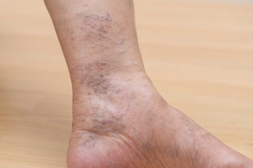
Corona Phlebectatica: Fan-Shaped Clusters at the Ankles
Corona phlebectatica is a strong indication of venous insufficiency of the saphenous veins. This presentation involves a large cluster of spider veins developing near the ankle in a distinctive fan-shaped or radial arrangement. The term "corona" refers to the crown-like pattern these vessels create. This specific configuration reliably indicates chronic venous insufficiency in deeper veins.
When Dr. Prevosti identifies corona phlebectatica during an examination, he recommends venous duplex ultrasound to map the deeper venous system. This spider vein pattern doesn't develop randomly; it results from sustained increased pressure in the venous system that forces blood into the tiny surface vessels. Treating only the visible spider veins without addressing the underlying venous insufficiency typically leads to rapid recurrence and continued symptoms.
Leg Symptoms That Suggest Venous Problems
Specific leg symptoms accompanying spider veins suggest underlying venous disorders. Leg aching, heaviness or tiredness that worsens throughout the day indicates blood is pooling in the lower extremities due to failing vein valves. Swelling that increases as the day progresses indicates that the venous system is struggling to return fluid from the legs to the heart efficiently.
Night cramps and restless legs frequently accompany venous insufficiency. While the exact mechanism isn't completely understood, the pooled, deoxygenated blood in distended veins appears to irritate surrounding nerves and muscles. Patients report that these symptoms often improve significantly after treating the underlying venous disorder.
Skin Changes That Raise Concerns
Skin changes near spider veins raise concerns about the progression of venous insufficiency. Redness, dryness, itching, discoloration or thickening of the skin in the lower calf or around the ankles indicates chronic inflammation from sustained venous pressure. Brown discoloration, known as hemosiderin staining or hyperpigmentation, develops when red blood cells leak from pressurized veins into the surrounding tissue, leaving iron deposits that permanently stain the skin.
Rapid Development or Spreading Patterns
The development of visible varicose veins alongside spreading spider veins strongly suggests progressive venous insufficiency. When spider veins appear rapidly or continue to spread despite conservative measures, evaluation becomes essential to identify any underlying valve dysfunction that may be driving their formation.
Dr. Prevosti's Diagnostic Approach
Patients with asymptomatic spider veins that are of pure cosmetic concern can come in for a “free screening,” where Dr. Prevosti verifies there are no signs or symptoms of venous insufficiency. If the evaluation confirms no need for a deeper evaluation, those patients can proceed with sclerotherapy.
Determining whether spider veins indicate an underlying problem requires specialized expertise and appropriate diagnostic technology. Dr. Prevosti's comprehensive evaluation process at Prevosti Vein Center provides patients with accurate answers about their venous health.
For patients with evidence of more than just cosmetic spider veins, the evaluation begins with a detailed discussion of your symptoms, medical history and family history of venous disorders. Dr. Prevosti asks specific questions about when spider veins first appeared, whether they've increased in number or size, and what symptoms you experience throughout the day. This information helps him understand the pattern of your condition.
Physical examination follows, with careful assessment of spider vein patterns and locations. Dr. Prevosti looks for the corona phlebectatica configuration that indicates deeper venous insufficiency. He examines your legs for varicose veins, skin changes, swelling and other signs of venous disorders. His decades of cardiovascular experience enable him to recognize subtle findings that less experienced providers might overlook.
Venous Duplex Ultrasound
Venous duplex ultrasound examination represents the definitive diagnostic tool for evaluating venous function. This advanced ultrasound technology maps your venous system, showing which veins function normally and which demonstrate valve failure that allows backward blood flow.
Dr. Prevosti's credentials as an RPVI (Registered Physician Vascular Interpreter) and RPhS (Registered Phlebology Sonographer) demonstrate his advanced expertise in performing and interpreting vascular ultrasounds. These certifications require extensive testing, documented experience and ongoing education. His skill with ultrasound technology enables him to identify complex patterns of venous insufficiency and develop treatment plans that address the root causes of symptoms.
Treatment Implications Based on Your Diagnosis
The findings from your evaluation determine the appropriate treatment approach. Dr. Prevosti's 15 years of focusing exclusively on vein care mean he has extensive experience matching patients with the optimal treatment strategy.
Treatment for Purely Cosmetic Spider Veins
When spider veins develop in isolation without underlying venous insufficiency, surface sclerotherapy effectively eliminates the visible vessels. Dr. Prevosti uses FDA-approved Asclera (polidocanol) injected directly into the spider veins through fine needles. The medication damages the cells lining the vein wall (the endothelium), causing the vein to close down. Your body gradually absorbs the collapsed vessels over weeks to months, and they fade from view.
Sclerotherapy sessions typically take approximately 20 minutes. Many patients require multiple treatments spaced one to three months apart for optimal results. Dr. Prevosti's thousands of sclerotherapy treatments provide him with the precision needed to achieve excellent outcomes while minimizing side effects. Patients are asked to wear compression stockings for three days after a sclerotherapy session. For patient convenience, appropriate compression stockings are available for purchase at the office. Normal, light daily activities can resume immediately after treatment with minimal restrictions.
Treatment When Venous Insufficiency Is Present
When diagnostic ultrasound reveals underlying venous insufficiency, treatment becomes more comprehensive. Addressing only the visible spider veins without correcting the deeper valve dysfunction that's driving their formation can result in less ideal results. The sustained pressure in the venous system continues forcing blood into surface vessels.
Dr. Prevosti's approach in these cases involves first treating the underlying venous insufficiency with minimally invasive procedures that seal the malfunctioning deeper veins, redirecting blood through healthy vessels and normalizing venous pressure. The surface veins are then treated.
Treatment options for venous insufficiency include:
- Radiofrequency ablation uses controlled thermal energy to seal damaged veins
- Endovenous laser ablation (EVLA) precisely targets malfunctioning veins with laser energy
- VenaSeal closure system uses a medical adhesive to permanently close problematic veins without heat
- Varithena foam therapy employs FDA-approved injectable foam to collapse and seal diseased veins
- Ultrasound-guided foam sclerotherapy addresses tributary veins and reticular veins (the slightly larger, bluish veins that often feed clusters of spider veins)
Once the root cause is corrected, surface sclerotherapy for the spider veins can begin.
Contact Prevosti Vein Center for Expert Evaluation and Treatment of Spider Veins
Even when spider veins appear purely cosmetic, evaluation provides valuable information about your venous health. Dr. Prevosti's examination can detect early venous insufficiency before it progresses to more advanced stages with skin damage or ulcers. Early intervention prevents complications and typically requires less extensive treatment than advanced cases.
If spider veins concern you or if you've noticed accompanying symptoms, contact Prevosti Vein Center to schedule your consultation. Call (470) 567-9047 or get in touch online today.
Take Back Your Comfort and Confidence With Prevosti Vein Center
Every patient at Prevosti Vein Center receives personal attention from Dr. Prevosti, a vein specialist with decades of experience in cardiovascular surgery. At Prevosti Vein Center, you will have access to the caliber of diagnostic and treatment approaches only available from a dedicated specialist.
Dr. Prevosti employs a compassionate and patient-centered approach that can reduce your symptoms and improve your comfort and daily activity level. His commitment to excellence in vein care has helped thousands of patients regain the confidence and freedom they need to pursue active lifestyles without the limitations of vein disorders.
Contact Prevosti Vein Center today to schedule an evaluation and discover how an experienced vein specialist can improve your quality of life.
